
Pathology reports that include genetic testing include technical words. Here are some of the more common terms you may see, with explanations in plain language.
Genetic tests look for changes in DNA that may help explain a health condition, guide your treatment, or provide information about risks that could be inherited within a family.
Some terms in your report describe the structure of genes, while others describe the types of changes that were found, or how the laboratory has interpreted those changes.
You can use this guide alongside your report to look up unfamiliar words as you read. If a term is not listed here, make a note of it and ask your doctor or a genetic specialist to explain it to you.
Use this explanation as a starting point, then discuss any questions or concerns with your doctor or genetic specialist.
These terms describe the structure of genes.
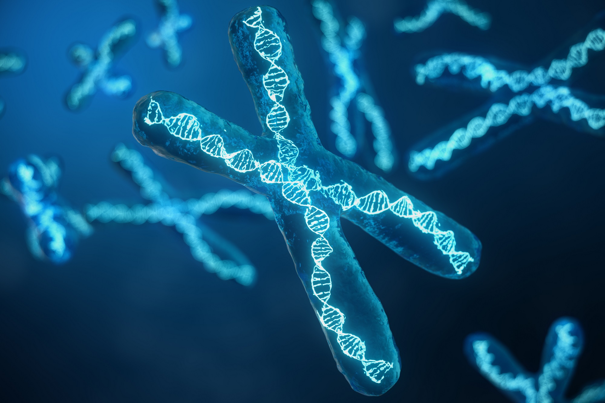
| Chromosomes | Chromosomes are structures found inside the nucleus of almost every cell in the body. They contain our DNA which is packaged in a highly organised way. In humans there are 46 chromosomes (23 pairs). One chromosome from each pair comes from the mother, and one from the father.
|
| DNA | DNA is the genetic material inside your cells. It carries the instructions for how your body grows, develops, and functions. |
| RNA | RNA is a translated version of DNA, that helps reveal the instructions stored in DNA. There are many different types of RNA. Messenger RNA (mRNA) is a temporary copy of a gene. It carries the DNA instructions to the cell machinery that builds proteins. |
| Gene | Genes are short sections of DNA. Genes tell the body how to grow, work and react to its surroundings. Genes do this by giving instructions to make proteins. Each gene carries the instructions for making a specific protein or carrying out a specific function in the body. |
| Adenine (A), Cytosine (C), Guanine (G), and Thymine (T). | Information in DNA is stored in the sequence of four chemical building blocks: adenine (A), cytosine (C), guanine (G) and thymine (T). These building blocks, called nucleotides, link together to form DNA. A,C,G and T are arranged in sequences that spell out specific instructions. They are sometimes called the Genetic Alphabet or DNA letters. |
| Allele | One version of a gene. Most people have two alleles of each gene — one inherited from each parent. |
| Exon | A part of a gene that contains active instructions used to build a protein. Genes usually contain multiple exons which are spliced together to make the final instructions for building the protein. |
| Intron | A part of a gene that does not contain instructions to make proteins and is cut out when exons are spliced together. |
| Promoter | A region of DNA that controls whether a gene is switched on or off. |
| Proteins | Proteins are the molecules that do the work in cells. They build cell structures, carry signals, control chemical reactions, transport substances, and help protect the body from infection. A protein is a large molecule made from a chain of smaller units called amino acids, linked together in a specific sequence. That sequence, determined by DNA, dictates how the protein folds, and the folded shape determines what the protein does. |
| Amino acid | A building block of proteins. Changes in DNA can alter the order of or how many amino acids are in a protein, which may affect how a protein works. |
| Structural variant | A larger-scale change to the structure of DNA, such as a section that is deleted, duplicated, inverted, or moved to a different location. |
| Genome | The complete set of genetic material in a person. |


A variant is any difference in DNA compared with the reference sequence. The reference sequence is a standardised comparison point for identifying and reporting genetic variants. Variants range from a single changed letter to large pieces of missing or extra DNA.
| Variant | A difference in DNA compared with the usual (reference) sequence. Variants may be harmless, or they may contribute to a health condition. |
| Insertion | Extra nucleotides added into a gene. |
| Deletion | A missing piece of DNA, ranging from a single letter to a large section of a gene. |
| Indel | A small insertion or deletion of DNA letters (a combination of both terms). |
| Copy number variant (CNV) | A section of DNA that is either missing (deleted) or present in extra copies (duplicated). |
| Missense variant | A change in a single DNA letter that causes a different amino acid to be used when building the protein. This may or may not affect how the protein works. |
| Nonsense variant | A change that creates an early “stop” signal in the DNA instructions, resulting in a protein that is shorter than normal. |
| Frameshift variant | An insertion or deletion that shifts how the DNA instructions are read, usually changing the protein significantly from that point onwards. |
| Splice site variant | A change that affects how sections of a gene are joined together during processing. This can disrupt the final protein that is produced. |
| Protein-truncating variant | Any change that shortens a protein. For example, a nonsense variant, a frameshift variant, or certain splice site variants. These variants usually lead to loss of normal protein function. |
| Sporadic gene changes | Sporadic gene changes occur by chance and are not inherited from either parent. They occur through natural DNA copying errors, environmental influences, or age related processes. Sporadic changes can be caused by de novo changes (present from conception) or by somatic changes (which occur later in life in only some cells, such as in a tumour). |
| De novo gene changes | A de novo gene change is a new genetic change that occurs for the first time in a person and is not inherited from either parent. It happens before or at conception and is present in every cell of the body. De novo changes can be inherited by the person’s children. |
| Gene fusion | A gene fusion occurs when two separate genes join because of a chromosomal rearrangement, forming a hybrid gene that can produce an abnormal protein. Gene fusions are common in cancer and can be important targets for treatment. |
| Translocation reciprocal, balanced, unbalanced | A translocation is when a piece of one chromosome breaks off and attaches to another chromosome, or when two chromosomes swap pieces. There are three main types of chromosomal translocations. A reciprocal translocation is where there is a two way exchange. Segments are swapped and no DNA is gained or lost only rearranged. Carriers are often healthy. A balanced translocation is where pieces of chromosomes break and reattach in new positions without any loss or gain of genetic material. People with balanced translocations are usually healthy but may have an increased chance of having pregnancies affected by unbalanced chromosome changes. An unbalanced translocation is a chromosome rearrangement where extra or missing genetic material is present because chromosome pieces have not been exchanged evenly. This can lead to health or developmental problems depending on which genes are affected. |
| Robertsonian translocation | A Robertsonian translocation is a special type that occurs only between chromosomes 13, 14, 15, 21 and 22. Carriers usually have 45 chromosomes but are healthy. There is an increased chance of unbalanced chromosome sets in offspring. This is the type involved in some cases of Down syndrome (e.g., 14;21 translocation). |
| Double strand DNA breaks | Double strand DNA breaks are serious forms of DNA damage where both strands of the DNA molecule are cut. They can be caused by natural cell processes, or external stresses such as radiation, chemicals, and infections. The body must repair them quickly. |
Your report may describe whether a variant was found in all your cells, or only in certain cells such as cancer cells.
| Germline | A genetic change that is present in all cells of the body. It was either inherited from a parent or arose very early in development, and it may be passed on to your children. |
| Somatic | Somatic changes are not inherited and cannot be passed on to children. They occur due to errors in DNA replication, exposure to environmental mutagens (compounds that damage DNA), or random variants that accumulate over time. Cells have a lifespan, they are constantly being renewed, and to do this they divide and replicate to grow more cells. Each time a cell divides, it copies its DNA and sometimes, small copying mistakes can occur. Most alterations are harmless, and some are repaired automatically by our cells but as we age our DNA repair mechanisms weaken. This means that some abnormal cells survive and multiply Sometimes this can causes cancer. |
| Zygosity | A description of whether a variant is present in one or both copies of a gene. |
Zygosity sub-types:
Laboratories use a standardised five-level scale to describe how likely a variant is to cause disease. This classification is based on scientific evidence and may be updated as new research becomes available.
| Benign | A change that is known not to cause disease. |
| Likely benign | A change that is very unlikely to cause disease, but there is not yet enough evidence to be completely certain. |
| Variant of uncertain significance (VUS) | A change where it is not yet known whether it causes disease. More research, or testing of other family members, may help to clarify its significance over time. |
| Likely pathogenic | A change that is very likely to cause disease, based on current evidence. |
| Pathogenic | A change that is known to cause disease. |
These terms describe how the test was done and how the results are described.
| Gene panel | A test that examines a selected group of genes at once. Panels are designed around genes known to be linked to a particular condition or group of conditions. |
| Sequencing | A laboratory method used to read the order of DNA letters in a gene or region of the genome. |
| Coverage | A measure of how completely each part of a gene or region was read during testing, and how many times each section was checked. |
| Reference sequence | The standard DNA sequence used as a comparison point when identifying and describing variants. |
Genetic tests use different laboratory methods to read, measure, or examine DNA. Each method has its own strengths and limitations, and laboratories often use more than one approach to provide the most accurate and complete result.
Sequencing is the process of reading the order of nucleotides (DNA letters) in a gene or region of the genome. There are several different sequencing methods, each suited to different purposes.
| Sequencing (DNA sequencing) | A method used to read the order of DNA letters in a gene or part of the genome. Sequencing can detect many types of variants, including single-letter changes, small insertions, and small deletions. |
| Next-generation sequencing (NGS) | A modern form of sequencing that reads millions of DNA fragments at the same time. NGS allows laboratories to test many genes together for example, as part of a gene panel, an exome test, or a whole-genome test. |
| Whole-exome sequencing (WES) | A test that reads all the exons (the coding parts of genes) across the genome. Most known disease-causing variants occur in exons, making WES a powerful tool for diagnosing genetic conditions. |
| Whole-genome sequencing (WGS) | A test that reads almost all of a person’s DNA, including exons, introns, and regions between genes. WGS can detect a wider range of variant types than other methods. For more on WGS see here. |
| Sanger sequencing | An older but highly accurate sequencing method. It is often used to confirm a variant found by NGS, or to examine a single gene region in detail. |
These techniques measure whether sections of DNA are missing (deletions) or present in extra copies (duplications). These changes are called copy number variants (CNVs).
| MLPA | Multiplex Ligation-dependent Probe Amplification. A targeted method used to detect deletions or duplications in specific genes or exons. MLPA is often used when a particular region is already suspected. |
| Microarray (chromosomal microarray / CMA) | A genome-wide method that can detect larger deletions, duplications, and some structural changes across the entire genome at once. |
These techniques look at the chromosomes directly, rather than reading individual DNA letters. They are used to detect large-scale changes.
| FISH (Fluorescence In Situ Hybridisation) | A technique that uses fluorescent probes to examine specific chromosomes or genes under a microscope. FISH can detect large deletions, duplications, and rearrangements in targeted regions. |
| Karyotyping | A test that examines the number and overall structure of chromosomes. Karyotyping can detect large-scale changes such as extra chromosomes, missing chromosomes or major rearrangements. For more on karyotyping see here. |


Some genetic conditions require specific techniques that go beyond standard DNA sequencing.
| RNA analysis (transcript analysis) | A method that examines RNA rather than DNA, to understand how a variant affects gene activity or how sections of a gene are joined together (splicing). This can help determine the significance of uncertain variants. |
| Methylation testing | A technique that measures chemical marks on DNA that control whether genes are switched on or off. It is used for conditions where abnormal methylation affects gene function, such as certain imprinting disorders. |
Your report may also include terms that describe how the test was performed or how the data was processed.
| Bioinformatics analysis | Computer-based methods used to assemble sequencing data, identify variants, and compare them with reference databases. This step is essential for interpreting the results of modern genetic tests. |